Veterinary Technicians, we've announced two new dates this Fall for our dental radiology lecture and hands-on lab! Seats are filing fast, sign up today!
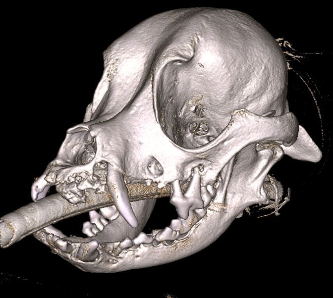
Oral imaging is an essential diagnostic tool that allows the doctor to visualize the bone under the gums to determine the extent of bone loss in areas of disease. The two types of imaging used in veterinary dentistry are dental radiographs and cone beam computed tomography (CBCT). Dental radiography uses a small film or sensor placed in the mouth. An x-ray beam passes through hard and soft tissue creating an image on the film which is transmitted back to a computer for viewing. CBCT also utilizes x-ray beams to produce images. The images produced with conventional dental radiographs are in 2-dimensions whereas the images produced by a CBCT can be used to create a 3-dimensional image of the tooth and bone structures, resulting in a more detailed view of the oral structures.